The New STED technique enables deep-tissue imaging and reveals the subcellular dynamics of neurons.
Researchers have created a new microscopy technique to capture 3D superresolution images from subcellular structures up to 100 microns below the surface of biological tissue. This includes the brain. The method allows scientists to see deeper into the brain and could reveal subtle changes in neurons that happen over time or due to disease.
This new approach extends stimulated emission depletion microscopy (STED), a groundbreaking technique that allows nanoscale resolution and overcomes the traditional diffraction limit in optical microscopes. This superresolution imaging technique was developed by Stefan Hell, who won the Nobel Prize in Chemistry in 2014.
The Optica journal of high-impact research by The Optical Society (OSA) describes how the researchers used their new STED microscope to photograph, in superresolution, dendritic spines deep within the brain of a live mouse. The dendritic spines, tiny protrusions found on the dendritic branch of neurons, receive synaptic inputs. They are crucial in neuronal activity.
Joerg Bewersdorf, Yale School of Medicine’s research team leader, said that the microscope was the first to achieve 3D STED superresolution deep within a living animal. Bewersdorf stated that deep-tissue imaging technology has allowed researchers to see subcellular structures and dynamics within their native tissue environment. This is crucial for understanding biological phenomena completely, both for biomedical research and pharmaceutical development.
The 3D-2PE–researchers used the STED microscope to view the brain of a live mouse. Zooming in on a portion of a dendrite shows an individual spine’s 3D structure and details. Credit: Joerg Bendersdorf, Yale School of Medicine
Going deeper
Conventional STED microscopy can be used to image cell culture specimens. It is more difficult to use the technique to image dense tissue or living animals, significantly when the superresolution benefits of STED can be extended to the third dimension for 3D STED. This is because optically thick tissues prevent light from reaching deep into the tissue and adequately focusing, limiting the superresolution capabilities of the STED microscope.
The researchers used STED microscopy, two-photon excitation (2PE), and adaptive optics to overcome this problem. Mary Grace M. Velasco was the first author of this paper. “2PE allows imaging deeper into tissue using near-infrared wavelengths instead of visible light.” “Infrared light has a lower rate of scattering, and is, therefore, better able penetrate deeper into tissue.”
The researchers also included adaptive optics in their system. Velasco stated that adaptive optics correct distortions in the light’s shape, i.e., optical aberrations, which can arise from imaging through and within the tissue. The adaptive element modifies light wavefronts in imaging in a way opposite to the specimen’s tissue. The adaptive component cancels out the aberrations in the tissue and creates ideal imaging conditions which allow for the STED superresolution capabilities of all three dimensions.
The brain changes you see
Researchers tested 3D-2PESTED by first imaging well-characterized structures on cultured cells using a cover slip. 3D-2PE–STED was able to resolve volumes ten times faster than 2PE. Their microscope detected the DNA distribution in mouse skin cells’ nuclei much better than a two-photon microscope.
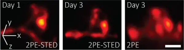
The researchers then used their 3D-2PE–STED microscope to photograph the brain of a live mouse. They focused on a specific part of the dendrite to resolve the 3D structure of individual spinal bones. Two days later, they imaged the same area again and found that the spine structure had changed. Researchers did not notice any changes in the neuron structure or behavior in the mice that could indicate damage to the imaging. They plan to continue their research.
Velasco stated that the dendritic spines are so tiny that it is hard to see their exact 3D shape without superresolution. “3D-2PE–STED allows us to see these changes not only in the brain’s superficial layers but also deep within, where there are more interesting connections.”


Comments